The Golden Hour Concept
The "golden hour" is one of the most important concepts in trauma surgery. It refers to the first 60 minutes after a major injury — the window during which rapid, systematic intervention has the greatest impact on survival and long-term outcome.
India accounts for approximately 11% of all road accident deaths worldwide — over 150,000 fatalities annually. A significant proportion of these deaths are preventable with timely trauma care. Road traffic accidents (RTAs) are the leading cause of polytrauma in India, and fractures — particularly of the long bones, pelvis, and spine — are almost universal in high-energy RTA victims.
Common RTA Mechanisms
- Two-wheeler vs. vehicle collision
- Pedestrian struck by vehicle
- Head-on collision at speed
- Fall from height / rollover
- Crush injuries under vehicle
Most Common Fractures in RTA
- Femur — massive blood loss risk
- Tibia/fibula — high open fracture rate
- Pelvis — life-threatening haemorrhage
- Spine — risk of paralysis
- Skull/facial bones
- Wrist (Colles') — outstretched hand injury
At the Accident Scene — What Bystanders Should Do
In India, the bystander often reaches a trauma victim before any ambulance. Simple actions in the first minutes can be life-saving. The Supreme Court of India has clarified that Good Samaritans helping accident victims are legally protected.
Call for Help — 108 Ambulance
Call 108 (national ambulance service) immediately. Give your location, the number of victims, and describe visible injuries. Stay on the line for instructions. Do not hang up.
Do NOT Move the Patient Unless in Immediate Danger
Moving a trauma victim with a spinal injury without neck support can cause permanent paralysis. Only move the patient if they are in a burning vehicle or submerged in water. Otherwise, keep them still until trained help arrives.
Control Visible Bleeding
Apply firm direct pressure with a clean cloth to bleeding wounds. Do not remove the cloth once applied — add more on top. Do not use a tourniquet unless you have been trained to do so and the bleeding is life-threatening from a limb.
Keep the Victim Warm and Reassured
Trauma victims go into shock and lose body heat rapidly. Cover them with any available cloth. Speak calmly to keep them conscious. Note the time of the accident — this information is crucial for the trauma team.
Do NOT Give Food, Water, or Medication
An unconscious or semi-conscious patient may aspirate. The patient may need emergency surgery — an empty stomach is essential for anaesthesia. Do not give any oral substances unless instructed by a trained dispatcher.
ATLS Primary Survey — What Happens in the ER
When a polytrauma patient arrives at a trauma centre, the team follows the ATLS (Advanced Trauma Life Support) protocol — a systematic, priority-based assessment that addresses life threats in order:
Airway with C-Spine Control
Is the patient breathing? Is the airway clear? The cervical spine is immobilised with a collar until injury is ruled out. Intubation is performed if the patient cannot maintain their airway.
Breathing and Ventilation
Check for pneumothorax (collapsed lung), haemothorax (blood in chest), or flail chest. Oxygen is administered. Chest drains inserted if needed. This is assessed before any limb fracture.
Circulation and Haemorrhage Control
Two large IV lines placed, blood drawn for cross-matching. Massive haemorrhage protocol activated if signs of shock. Pelvis and femur fractures are the most common source of hidden haemorrhage — a pelvic binder is applied for suspected pelvic ring injuries.
Disability — Neurological Status
GCS (Glasgow Coma Scale) assessed. Pupil reaction checked. Any limb weakness, numbness, or loss of bladder/bowel control noted — these suggest spinal cord involvement.
Exposure and Environment
All clothing removed to identify every injury. Kept warm to prevent hypothermia. A full secondary survey (head-to-toe examination) follows once life threats are controlled.
Common Fracture Patterns in Road Accidents
Femoral Shaft Fracture
A closed femur fracture can cause 1–2 litres of hidden blood loss into the thigh. Emergency management: skin traction to reduce blood loss. Definitive: intramedullary nail (IMN) — the gold standard, allowing early weight bearing.
Pelvic Ring Injury
Unstable pelvic fractures (APC-III, VS type) can cause life-threatening haemorrhage of 3–5 litres. Pelvic binder is applied in the ER, followed by external fixation or angioembolisation. Definitive ORIF when stable.
Tibial Shaft Fracture
Most common long bone fracture in India. High risk of open injury (Gustilo classification). Intramedullary nailing preferred. Compartment syndrome must be watched for in the first 24–48 hours.
Spinal Fracture
Thoracolumbar junction (T12–L1) most common. Neurological status assessed with ASIA scale. Unstable fractures or those with neurological deficit require surgical stabilisation — typically pedicle screw fixation within 24–72 hours.
Clavicle & Shoulder
Very common in two-wheeler accidents. Most clavicle fractures heal with sling + physiotherapy. Severely displaced fractures or those with neurovascular compromise need surgical plating.
Wrist Fractures (Colles')
Classic fall-on-outstretched-hand (FOOSH) injury. Undisplaced — plaster cast for 6 weeks. Displaced/unstable — closed reduction + K-wires or volar plating (ORIF).
Open Fractures — A Limb-Threatening Emergency
An open (compound) fracture — where bone penetrates the skin — is a surgical emergency. Contamination of bone with road debris, soil, and bacteria makes infection (osteomyelitis) a catastrophic risk if not treated urgently.
Gustilo-Anderson Classification
- Type I — wound <1 cm, minimal contamination
- Type II — wound 1–10 cm, moderate soft tissue damage
- Type IIIA — extensive soft tissue, adequate bone coverage
- Type IIIB — extensive periosteal stripping, needs flap cover
- Type IIIC — arterial injury requiring repair (amputation risk)
Emergency Management
- Sterile saline-soaked dressing — cover wound, don't scrub
- IV antibiotics within 1 hour
- Tetanus toxoid (if not up to date)
- Emergency surgical washout and debridement
- Temporary external fixator for stabilisation
- Definitive fixation once wound is clean (48–72h)
Damage Control Orthopaedics (DCO)
In a severely injured polytrauma patient, attempting complex definitive fixation of all fractures in the first surgery can overwhelm the body — a phenomenon called the "second hit" that dramatically increases mortality.
Damage Control Orthopaedics (DCO) is the principle of doing only the minimum necessary in the first surgery to save the limb and control haemorrhage, then performing definitive fixation 48–72 hours later when the patient has been resuscitated and stabilised.
Stage 1 — Emergency (0–24h)
- Pelvic binder / external fixator
- Fasciotomy for compartment syndrome
- Temporary external fixation of long bones
- Vascular repair if arterial injury
- ICU resuscitation: transfusion, warming
Stage 2 — Definitive (48–120h)
- Intramedullary nailing of femur/tibia
- ORIF of pelvis/acetabulum
- Spinal stabilisation
- Soft tissue reconstruction / flap cover
- Patellar/ankle/wrist fixation
Complications to Watch For
Compartment Syndrome
Pressure builds within the closed muscle compartments, cutting off blood supply. Classic signs: severe pain out of proportion to injury, pain on passive stretch of fingers/toes, tense swelling. This is a surgical emergency — fasciotomy must be performed within hours to prevent permanent muscle death and contracture.
Fat Embolism Syndrome (FES)
Fat droplets from fractured bone marrow enter the bloodstream, causing respiratory failure, neurological changes, and a petechial rash. Most common after femur and pelvic fractures. Early stabilisation of long bone fractures is the best prevention.
Deep Vein Thrombosis (DVT) & Pulmonary Embolism
Immobility after fracture greatly increases clot risk. All trauma patients receive DVT prophylaxis — low-molecular-weight heparin, compression stockings, and early mobilisation. PE remains a leading cause of preventable trauma death.
Non-Union and Malunion
A fracture that fails to heal (non-union) or heals in a poor position (malunion) may require revision surgery with bone grafting. Risk factors include open fractures, smoking, diabetes, infection, and inadequate fixation.
Rehabilitation After Trauma Fractures
Surgery fixes the fracture — rehabilitation restores the person. A structured physio programme is as important as the operation itself.
Week 1–2 — ICU / Ward Phase
Chest physiotherapy, breathing exercises, and limb exercises to prevent stiffness. Sitting up in bed, transfers to chair. DVT prevention is a priority.
Week 3–6 — Early Mobilisation
Weight-bearing status depends on fracture type and fixation. Partial weight bearing with crutches for most lower limb fractures. Active range-of-motion exercises for all adjacent joints.
Month 2–3 — Progressive Loading
Full weight bearing as X-rays show callus formation. Strengthening exercises. Hydrotherapy if available. Scar management for soft tissue injuries. Psychological support — PTSD is common after RTAs.
Month 3–6 — Return to Function
Return to driving, work, and daily activities. Sports rehabilitation for younger patients. Final X-ray at 6 months. Hardware removal (if planned) after full union — typically at 12–18 months for tibial nails.
Suffered a Fracture in an Accident?
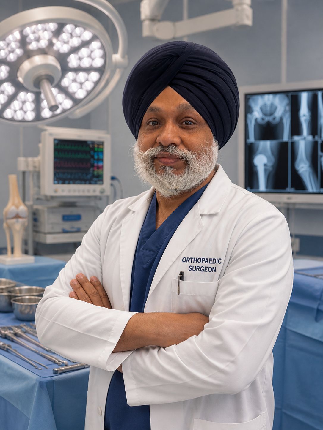
Dr. Maninder Singh provides comprehensive trauma orthopaedic care — from emergency fracture fixation to full rehabilitation. Early consultation ensures the best possible outcome for your recovery.